Sovereign Tier

LeptonX-Ray Vision

See what's yours to see.

Your scans. Your measurements. Your understanding.
The first on-device imaging intelligence platform that puts 3D tumor reconstruction and quantitative tracking entirely in the patient's hands.

Scroll
4,463Named Anatomical Structures
7Complete Body Systems
0Data Leaves Your Device
1Voice Command to Navigate
The Knowledge Gap

You paid for the scan.
You deserve more than a paragraph.

Today, a cancer patient receives a radiology report — a wall of jargon, a single longest-diameter measurement, and perhaps a flat grayscale image on a portal they don't control. That's the entirety of what most patients ever see of their own imaging.

The data inside your MRI is extraordinarily rich — volumetric, spatial, dimensional. But by the time it reaches you, it has been compressed into a sentence. You are given a caption when you own the photograph.

LeptonX-Ray Vision restores what was always yours. It takes your DICOM imaging data, reconstructs it in three-dimensional space, segments your anatomy with research-grade AI, and lets you see — truly see — what is happening inside your own body. On your device. Under your control. With no data leaving your hands.

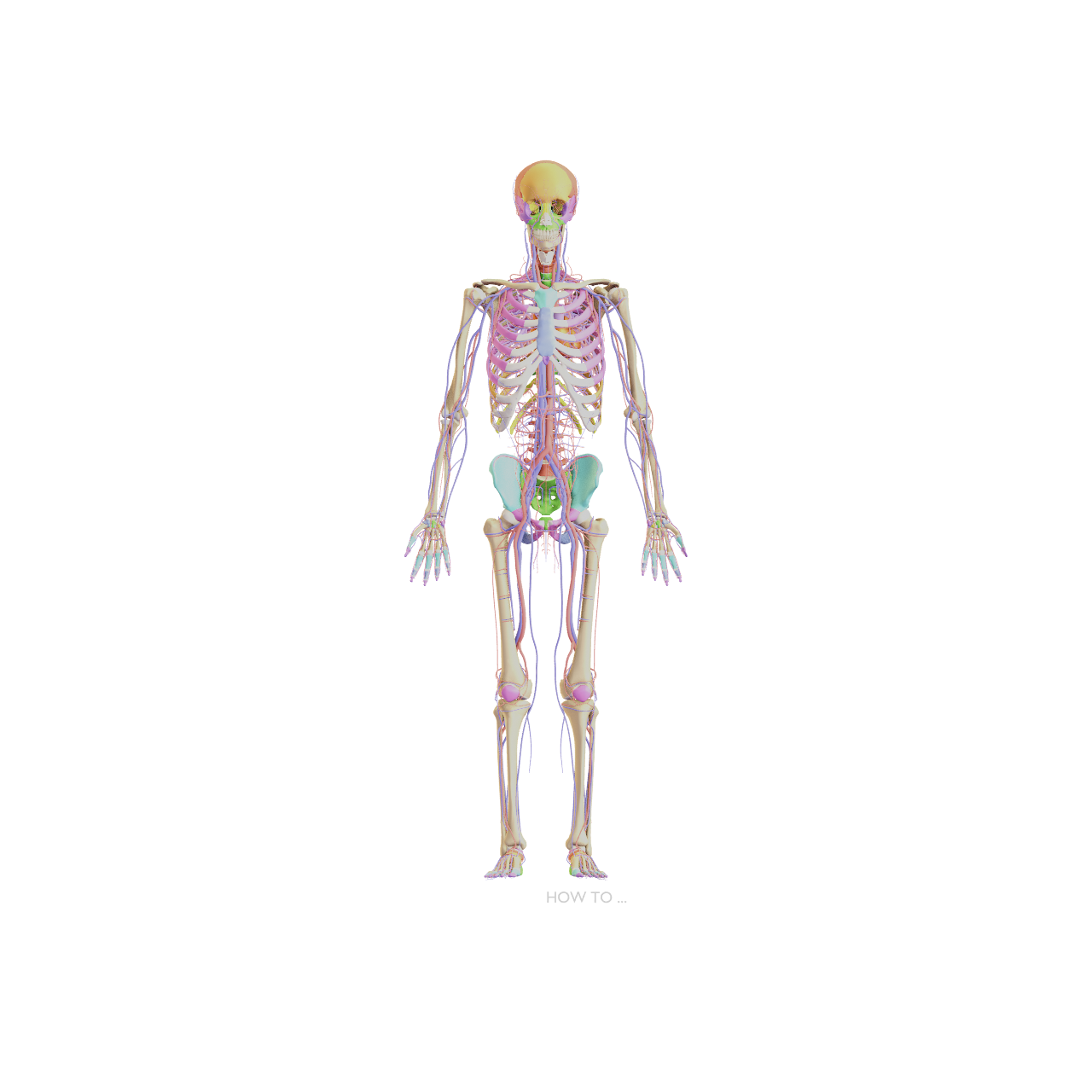
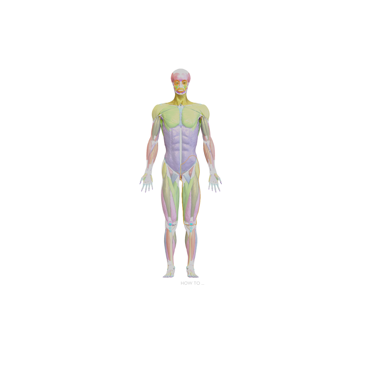
Skeletal & Circulatory Intelligence

Physicians spend a decade learning what lives inside you.
You deserve that same clarity.

Every time a doctor delivers news about your body — a diagnosis, a finding, a recommended procedure — there is an invisible translation layer between their knowledge and yours. You receive words where you need images. You receive abstractions where you need structure.

Maya's Anatomical Intelligence closes that gap permanently. When your physician tells you there is a finding near your iliac artery, Maya doesn't just explain it — she takes you there. The 3D model responds to your voice, flies to the exact structure, and surfaces everything your health record says about it.

This is not information retrieval. This is comprehension — personalized, private, and immediate.

Powered by First Particles™ — decomposing every medical concept to its most fundamental truth, then reconstructing it in language that reaches you.
Full body skeleton with circulatory system
Full body muscular system
Muscular System · 650+ Muscles Mapped

Every fiber accountable.
Every movement explainable.

Maya maps muscular anatomy directly to surgical reports, physical therapy notes, and imaging findings from your health record. When your orthopedist documents a rotator cuff finding, Maya shows you exactly where it is — in three dimensions.

650+ named muscles. Every attachment, every action, every clinical implication — cross-referenced against what your doctors have documented about your specific body.

Powered by First Particles™ — decomposing every medical concept to its most fundamental truth, then reconstructing it in language that reaches you.
How It Works

From scan to understanding

01

Your Data, Your Device

Connect via secure FHIR integration, or load DICOM files from a CD, USB, or portal download. Your raw imaging data arrives on your device and stays there.

02

AI Segmentation

Research-grade neural networks analyze your scan entirely on-device. They identify organs, vessels, bones, and lesions, producing precise three-dimensional masks of every relevant structure.

03

3D Reconstruction

Segmented structures become high-fidelity polygon meshes — smoothed, measured, and rendered against anatomically accurate organ models.

04

Quantitative Intelligence

Volume. Longest diameter. Distance to critical structures. Every number derived from your scan, computed independently, tracked across every scan you've ever had.

05

Maya Walks You Through It

Maya narrates what you're seeing in plain language — guiding you through the 3D scene, explaining measurements, and highlighting changes from previous scans.

06

Longitudinal Tracking

Every scan session is versioned. Volume deltas, dimensional trends, and treatment response — tracked over months and years with clinical precision.

What You See

Your body, rendered with fidelity

Tumor-to-Organ Spatial Mapping

See exactly where a mass sits relative to the organ it occupies — its proximity to vessels and ducts, its relationship to surgical margins. Not described. Shown.

Volumetric Measurement

True 3D volume computation captures irregular shapes, asymmetric growth, and response patterns that flat measurements miss entirely.

Serial Comparison

Overlay meshes from different timepoints. See growth and shrinkage color-coded in three dimensions — not from a number, from a shape.

Voice-Guided Navigation

Maya narrates the reconstruction — identifying structures, reading measurements, comparing to prior scans in language you understand.

Measurement Callouts

Dimensional annotations and margin measurements rendered directly in the 3D scene — spatial information anchored to anatomy.

Independent Verification

Run your own quantitative analysis. Arrive at your appointments with understanding, not just anxiety.

Female Pelvic Floor · 3D Interactive

Structures hidden from view.
Made visible at last.

The female pelvic floor — one of the most clinically significant and least understood regions of the body. Maya renders it in full 3D, navigable by voice, cross-referenced against your health record.

Model: University of Dundee, CAHID · Sketchfab
Pulmonary System · Live 3D

The lungs, alive in your hands.

Rotate, zoom, and inspect. When your pulmonologist documents a finding, Maya maps it here — in three dimensions, in plain language.

Model: Ebers · Sketchfab
Precision Commitment

We publish our accuracy.
Every number, verified.

A beautiful 3D rendering of an inaccurate segmentation is not a medical tool — it's a liability. LeptonX-Ray Vision is held to the LeptonX Imaging Fidelity Standard, validated against expert-annotated ground-truth datasets.

Imaging Fidelity Standard

Every segmentation model is validated against the Medical Segmentation Decathlon and published clinical datasets. We report our error margins. We do not ship capability without a published accuracy number.

Volumetric ErrorPublished per organ
Dimensional Accuracymm deviation reported
Surface FidelityHausdorff distance
ReproducibilityRepeat-run variance

We lead with precision, not with flair. If we can't prove the number, we don't show the number.

Sovereign by Architecture

LeptonX-Ray Vision does not upload your imaging data to a cloud. It does not send your scans through a third-party API. Every computation — segmentation, mesh generation, measurement, narration — executes on your device.

This is not a privacy policy. It is an architectural fact. There is no server to breach because there is no server.

Zero cloud dependency
Zero PHI transmission
Zero third-party data access
Zero custody of your medical imaging
It was always your body.
It was always your scan.
It was always your right to understand it.
First Particles™ — LeptonX Health Intelligence
Available at the Sovereign Tier

See what's yours to see.

LeptonX-Ray Vision is in active development as part of the LeptonX Sovereign platform.

LeptonX-Ray Vision is a patient comprehension and self-advocacy tool. It is not a diagnostic device and does not provide medical diagnoses, treatment recommendations, or clinical decision support. All imaging analysis should be discussed with your qualified healthcare provider.